Microscopy Facility
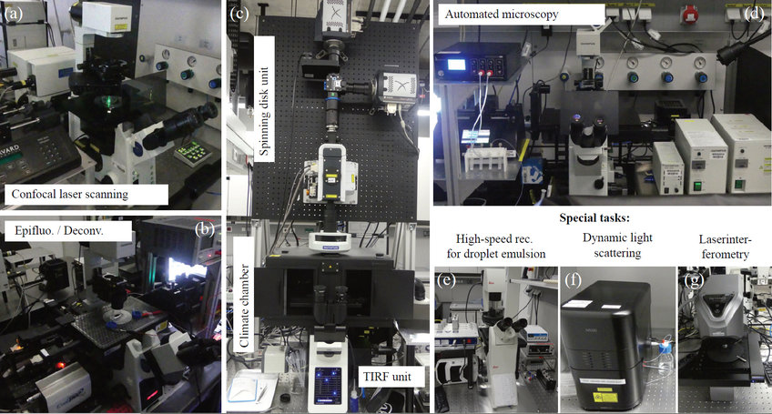
The microscopy facility of the MPI DS combines a set of advanced lightand laser based microscopes. It is operated by the LFPB department (Bodenschatz), but is open to users from the other departments as well as external users from the neighboring MPINAT. The current focus of the biophysical subgroups within the LFPB department are quantitative and synthetic biology. Therefore, the selection of the microscopes applies techniques of life cell imaging aided by sensitive cameras/detectors (see figure below). The backbone of the facility are the confocal laser scanning microscopes (Olympus FV1000) and a confocal spinning disk microscope (Olympus), which serve for a broad range of experiments, such as single cell stimulation and fast 3D imaging of migrating Dictyostelium discoideum. These are complemented by a high precision light microscopy set-up (Deltavision), which achieves its z-resolution by deconvolution of epifluorescence z-stacks.
After substantially enlarging the facility in the period 2016-2019, we now finalized the necessary upgrades and programming of the microscopes as well as the attached auxiliary hardware. Furthermore, we recently integrated multiple light and fluorescence microscopes of the now Emeritus group of Gregor Eichele. The different set ups allow for a variety of experiments such as large scale and long time unsupervised recordings, high speed video capture, dynamic light scattering and surface roughness measurements. As in the previous period, the service of the microscopy facility also comprises the training of new users and the education of apprentices.

microscope (Olympus FV1000). (b) High precision epifluorescence (deconvolution
enhanced) microscopy (Deltavision). (c) Dual set-up consisting of a spinning disk unit
and a Fluorescence/TIRF unit (Olympus BX63/IX83). (d) Fully automated microscopy
with large scale recording and additional control of auxiliary devices. (e-g) Set-ups
designed for special tasks.
