Sleeping sickness parasite masters three different swimming modes
Scientists from Germany and Switzerland decipher the precise patterns of motion of the parasite Trypanosoma.
The causative agent of African sleeping sickness, annually responsible for several thousands of deaths in Africa and South America, is a motile cell: it propels itself through its host’s bloodstream until – in the last stage of the disease – it overcomes the blood-brain-barrier and penetrates its victim’s brain. In order to fight this deadly disease, scientists are trying to understand the parasite’s exact patterns of movement. Researchers from the Max Planck Institute for Dynamics and Self-Organization (MPIDS) in Germany as well as from the Universities of Würzburg, Göttingen and Basel have now succeeded in identifying three different swimming modes. In addition, they were able to show for the first time, that these swimming modes correspond to the shape and stiffness of the parasite.

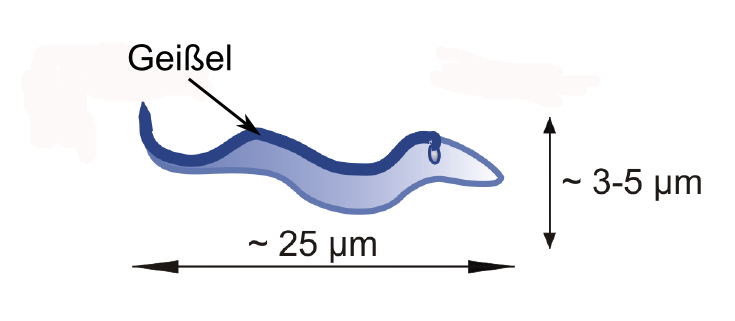
Cells that move independently are not uncommon: E. coli bacteria wrap their flagellum up into a tight bundle in order to propel themselves forward; sperm cells move by whipping their tails back and forth. In the case of Trypanosoma, parasites that cause African sleeping sickness, the exact motion-sequence is not yet known. The reason for this is their much more complicated “physique”: The flagellum, most probably the cell’s main motor, does not link to the cell body like a tail as is the case with sperm. Instead, it is connected to the cell on its entire length (see figure 1). What is for sure, however, is that Trypanosoma’s method of moving forward is extremely efficient: The parasites swim through their victim’s blood stream with velocities ranging from 20 to 40 μm/s.
The German-Swiss group of scientists was now able to show that Trypanosoma brucei brucei, the parasite that infects cattle, swims in three different swimming modes. Under a microscope the researchers tracked the exact paths chosen by the cells in a period of a few seconds within a nutrient solution and analyzed these statistically. As it turned out, the parasites could be divided into three groups: while some of them moved into the same direction for several seconds, others tumbled randomly every which way. “As a result, they hardly move forward at all”, Sravanti Uppaluri from the MPIDS describes this group. A third group switched between both swimming modes.
In order to study why a certain cell favours a certain “gait”, an even more precise – and especially quicker – look at Trypanosoma was necessary. “First, we had to track a cell for a certain period of time and identify its swimming mode”, explains Uppaluri who performed the challenging analysis. In a next step, the scientists took a closer look at the anatomical properties of the cells with the help of a high-speed camera, allowing for up to several thousands of images per second and a higher spatial resolution. By measuring the distance between both cell ends the researchers found that the directional swimmers have a stretched form indicating they are stiffer than their tumbling counterparts. The randomly tumbling Trypanosoma on the other hand proved to be rather more bent, pointing to a more flexible body.