Mechanics and dynamics of biological adhesion
Actin-based dynamics and periodic stimulation of Dictyostelium
The actin cytoskeleton, a dynamical, cross-linked biopolymer scaffold at the inner side of the plasma membrane is essential for many bio-mechanical properties of eukaryotic cells. Examples that rely on the rapid rearrangement of the actin cytoskeleton in response to external chemical cues are found in wound healing, in the morphogenetic development of an embryo, or in the metastatic spreading of cancer cells in the body (1). One of the most convenient and well studied eukaryotic model organisms is the social amoeba Dictyostelium discoideum, a single-celled eukaryote that exhibits chemotactic responses in gradients of extracellular cAMP (2).
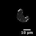
In a recent study, we have exposed single chemotactic Dictyostelium cells to well-controlled temporal stimuli of cAMP (3). For these experiments, we used the LimE-GFP labeled Dictyostelium strain (kindly provided by Günther Gerisch, see also (4)); LimE-GFP has previously been introduced as marker for filamentous actin (F-Actin) (5) and is shown in Fig.1. Short pulses and periodic pulse trains were generated using microfluidic flow photolysis, an approach that is based on the light-induced cleavage of caged cAMP in a microflow (6).

A confocal laser scanning image of Dictyostelium discoideum LimE-GFP: bright spots mark a high LimE-GFP concentration.
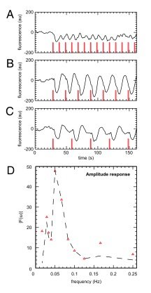
Single cell responses of the actin system to a short stimulus of cAMP included short, strongly damped responses of the actin machinery, as well as, slowly decaying, weakly damped oscillations. In a small subpopulation, even perpetual autonomous oscillations in the cortical actin density were observed. These findings suggest that the actin machinery of chemotactic Dictyostelium cells operates close to the onset of oscillations. Stimulation with periodic sequences of cAMP pulses confirmed these results (periodic forcing using a 405 nm 'uncaging' laser, periods ranging from 6 s up to 35 s). When monitoring the cytosolic and cortical actin response as a function of the stimulation frequency, we found a resonance at a stimulation period of around 20 s. Our findings can be explained in the framework of a generic model that captures the time-delay in the regulatory network of the actin system.

(A)-(C) Time series of the average cytosolic fluorescence intensities (black) of LimE–GFP cells responding to pulse trains with a period of (A) 10 s, (B) 20 s, and (C) 30 s. The laser stimulus is shown in red. In (D), the amplitude of the largest peaks in the frequency spectrum of the corresponding data sets are displayed as red triangles for each stimulus frequency. Furthermore, the amplitude at the stimulus frequency is shown (dashed line). A clear resonance is observed at around stimulation periods of T = 20 s. Adapted from (3).
By using cell lines that expressed GFP-tagged fusion proteins as well as knockouts of actin-binding proteins that enhance the disassembly of actin filaments, we work on confirming and extending our model.

